About FLAT PANEL BASED MOBILE C-ARM X-ray System
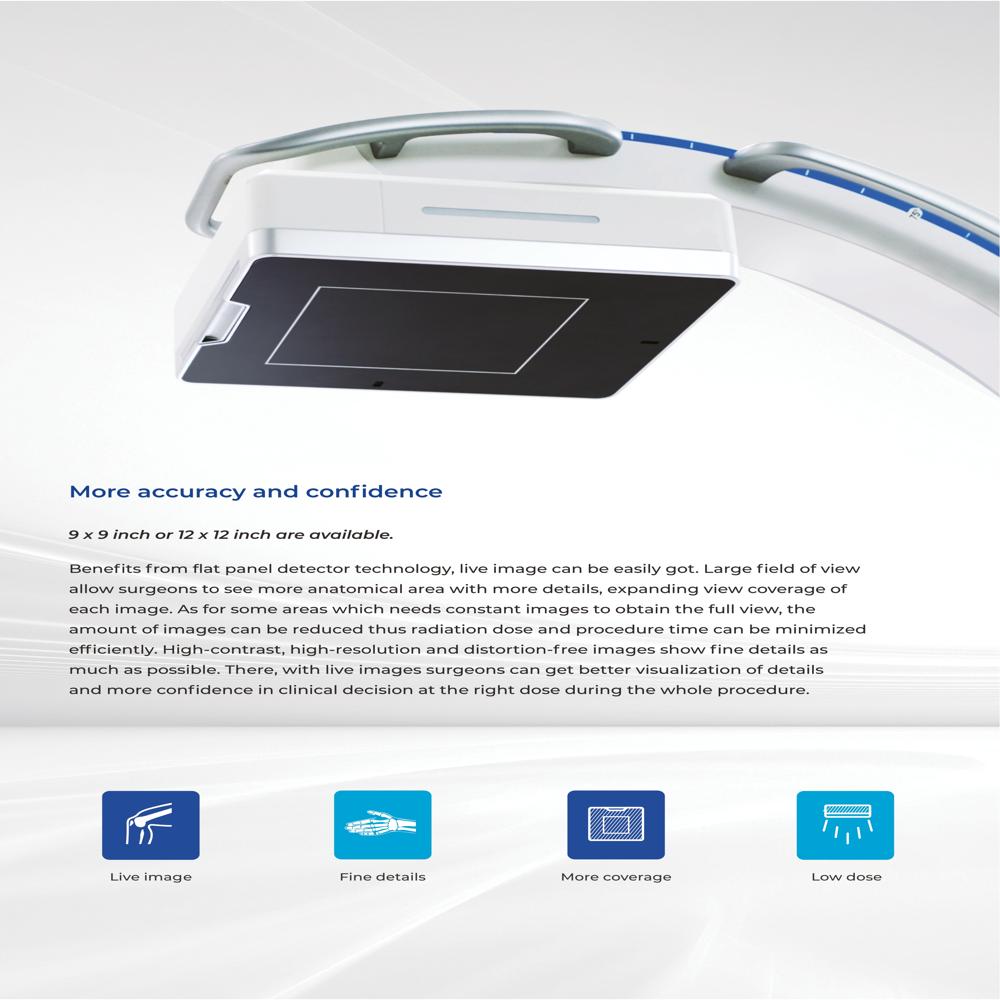
9 x 9 inch or 12 x 12 inch are available.
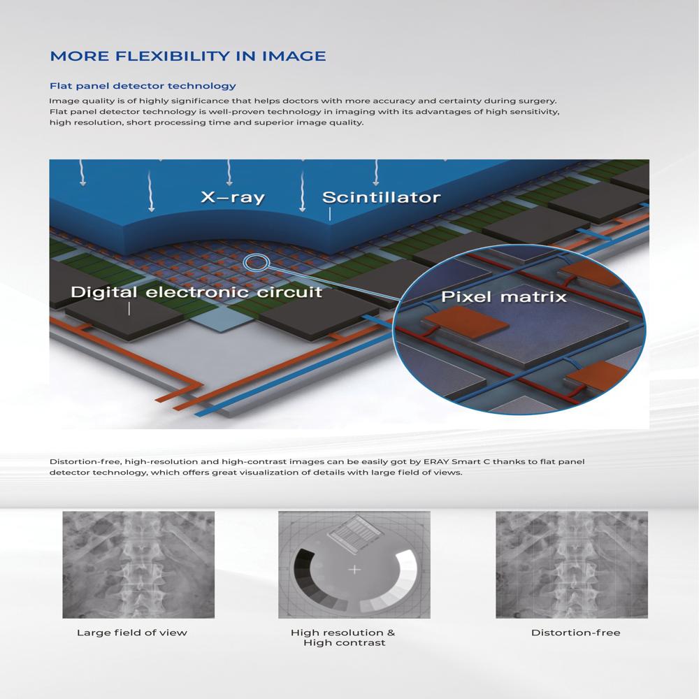
Benefits from flat panel detector technology, live image can be easily got. Large field of view allow surgeons to see more anatomical area with more details, expanding view coverage of each image. As for some areas which needs constant images to obtain the full view, the amount of images can be reduced thus radiation dose and procedure time can be minimized efficiently. High-contrast, high-resolution and distortion-free images show fine details as much as possible. There, with live images surgeons can get better visualization of details and more confidence in clinical decision at the right dose during the whole procedure.
Advanced Imaging TechnologyThis C-Arm X-ray system uses a high-resolution amorphous silicon flat panel detector that allows for near-instantaneous digital image capture. The advanced image processing algorithms provide enhanced clarity by reducing noise and improving visualization in both fluoroscopy and digital radiography modes. These features make it excellent for delicate intraoperative procedures requiring precision.
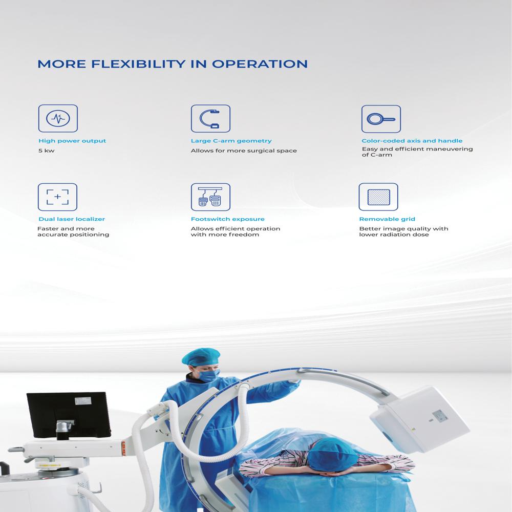
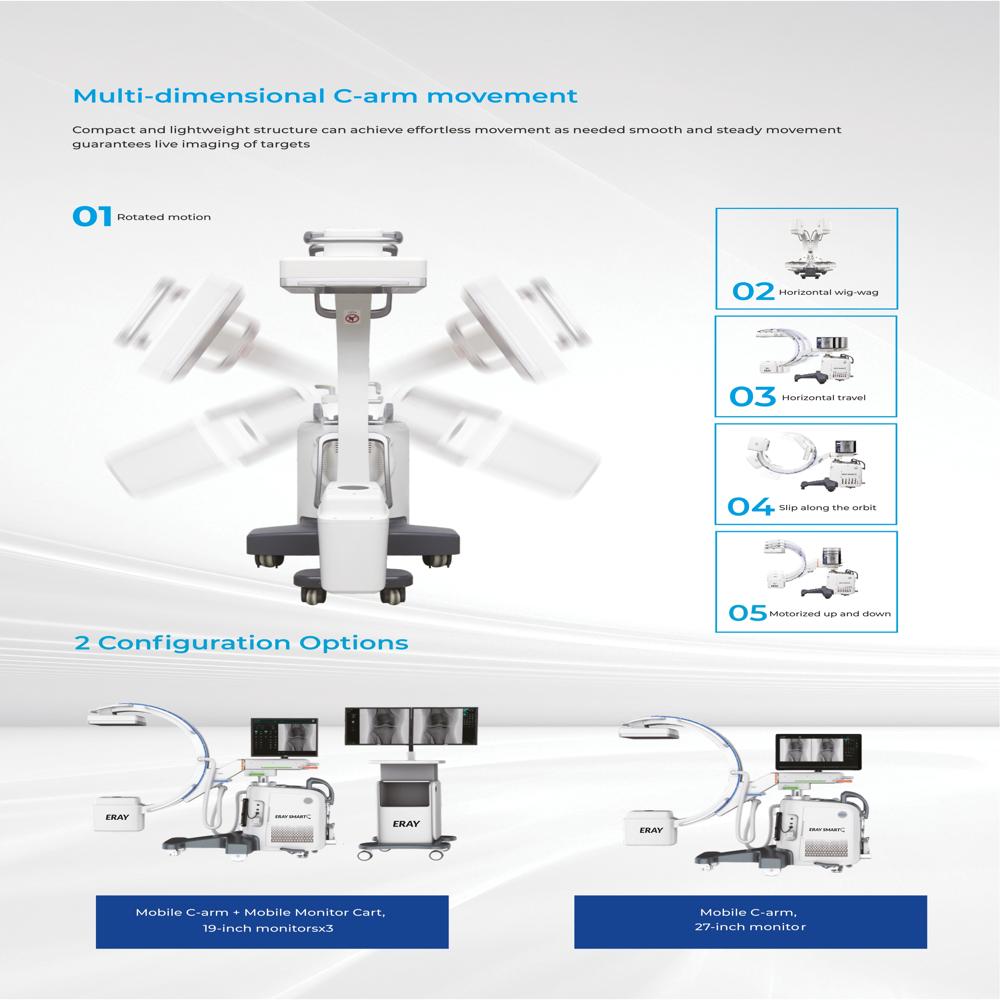
Flexible Movement and OperationMotorized vertical, horizontal, orbital, and swivel movements enable seamless positioning for various surgical and diagnostic procedures. The integrated laser beam aids precise centering, while adjustable arm and display height accommodate different operating environments, ensuring ergonomic use for health professionals.
Efficient Data Management and ConnectivityWith 500GB to 1TB internal storage and export options like DICOM, USB, CD/DVD, and network sharing, this system streamlines data management and sharing. The dual touchscreen monitors and multi-modality viewing features allow for easy image annotation, measurement, and archiving during procedures.
FAQ's of FLAT PANEL BASED MOBILE C-ARM X-ray System:
Q: How does the flat panel detector improve imaging quality?
A: The amorphous silicon digital flat panel detector (available in 21 x 21 cm and other sizes) delivers high-resolution images with advanced noise reduction and enhancement algorithms, ensuring precise visualization for diagnostic and surgical guidance.
Q: What types of procedures can this mobile C-Arm be used for?
A: This system is versatile and suitable for surgical imaging, orthopedics, emergency medicine, cardiology, and pain management, providing both fluoroscopy and digital radiography for intraoperative diagnosis and treatment guidance.
Q: When is the battery backup function useful?
A: The optional battery backup allows for short-term mobile operation, making it valuable during power outages, patient transport within hospitals, or in situations where a fixed power supply is unavailable.
Q: Where can the data from imaging procedures be exported?
A: Image data can be exported via DICOM for integration into hospital systems, or using USB devices, CD/DVDs, and network sharing, ensuring flexibility in data management and record-keeping.
Q: What makes the system easy to use during surgery?
A: Motorized movements (vertical, horizontal, orbital, swivel), an integrated laser for precise positioning, adjustable arm and display, and user-friendly touchscreen monitors streamline configuration and operation, minimizing disruptions during procedures.
Q: How does the device help control patient dose during imaging?
A: It includes pulse mode imaging and a Dose Area Product (DAP) meter, both designed to manage and monitor radiation exposure, prioritizing patient safety throughout the imaging process.


 Send Inquiry
 Send Inquiry
 Send SMS